Understanding Petroclival Meningioma
A Petroclival Meningioma is a rare type of skull base tumor that arises from the meninges near the junction of the petrous bone and the clivus — an area deep within the skull, close to the brainstem and cranial nerves. Although usually benign (non-cancerous), its location makes surgical treatment extremely challenging.
These tumors account for a small percentage of all meningiomas but can cause significant neurological symptoms due to their proximity to vital brain structures such as the brainstem, cranial nerves, and major blood vessels.
Common Symptoms
Because Petroclival Meningiomas grow slowly and are located deep within the skull, symptoms often appear gradually and vary based on the tumor’s size and direction of growth. Patients may experience:
- Double vision or facial numbness
- Hearing loss or ringing in the ear (tinnitus)
- Difficulty swallowing or speaking
- Weakness or imbalance while walking
- Persistent headaches
- Seizures (less common)
Diagnosis
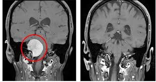
Accurate diagnosis requires detailed neuroimaging studies. MRI with contrast is the gold standard to identify the tumor’s size, location, and its relationship to nearby nerves and vessels. CT scans help assess involvement of surrounding bones at the skull base.
In some cases, angiography may be performed to map the tumor’s blood supply before surgery. A biopsy or surgical specimen confirms the diagnosis and determines whether the tumor is benign, atypical, or malignant.
Treatment Options
The goal of treatment is to achieve maximum safe tumor removal while preserving neurological function. Due to the deep location and proximity to critical structures, surgery is often complex and must be carefully planned. Common treatment approaches include:
- Microsurgical Resection: Performed using advanced skull base techniques to safely remove the tumor through minimally invasive routes.
- Neuronavigation & Intraoperative Neurophysiological Monitoring: Help ensure precision and protect critical nerves during surgery.
- Stereotactic Radiosurgery (Gamma Knife / CyberKnife): Ideal for small, residual, or recurrent tumors located near delicate structures.
- Fractionated Radiation Therapy: Used when complete removal is not feasible.
- Observation: For very small, slow-growing, or asymptomatic tumors under regular MRI follow-up.
Expert Petroclival Meningioma Care in Ahmedabad
At HCG Aastha Cancer Centre, Ahmedabad, Dr. Chirag Panchal, Consultant Neurosurgeon, offers advanced management for complex skull base tumors like Petroclival Meningiomas. With expertise in microneurosurgery, neuroendoscopy, and neuronavigation-guided skull base approaches, he ensures optimal tumor removal while minimizing neurological complications.
When to Seek Medical Attention
- Persistent or worsening headaches
- New onset of double vision or facial weakness
- Unexplained imbalance or dizziness
- Hearing loss or ringing in one ear
- Difficulty speaking or swallowing
Early diagnosis and expert management greatly improve outcomes in patients with Petroclival Meningioma. If you or someone you know is experiencing such symptoms, consult a skull base neurosurgeon promptly.
